About this treatment
Dental imaging rests on two examinations — the intraoral and the panoramic X-ray — both indispensable for uncovering hidden problems.
Intraoral X-ray
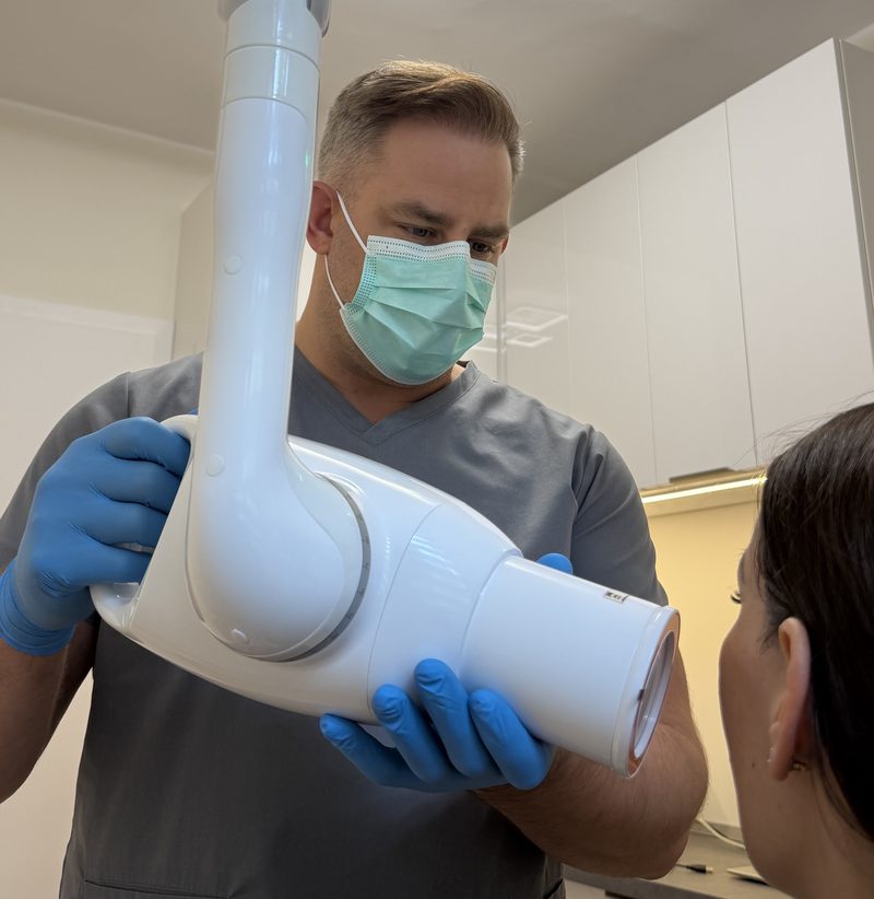
An intraoral X-ray is a small image showing one tooth and its root, which makes inflammation at the root tip or an inadequate filling visible. We also use it to check completed root fillings, so we can be certain the treatment was successful.
Panoramic X-ray
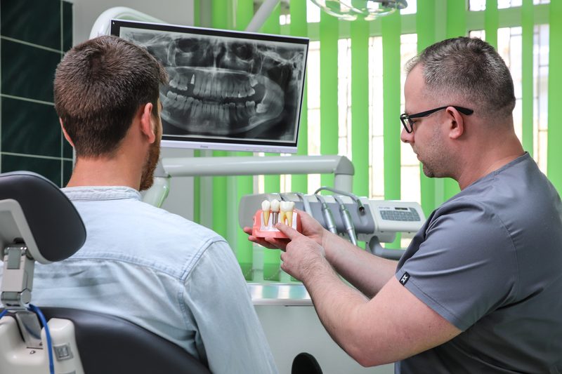
A panoramic X-ray shows all of the teeth in a single image, along with any decay, the jaws, the sinuses and the jaw joint. It is ideal for a thorough review of the dentition, for locating wisdom teeth and for focal infection screening.
The value of these images lies in making changes visible that the eye cannot see, allowing an early and accurate diagnosis and a precise, personalised treatment plan.